フォークリフトの仕事は、堅実に作業をこなす人にぴったりです。表舞台で働いている人を裏から支える事ができるのが良い点ですよね。目立つ仕事ではないものの、この仕事にもたくさんのやりがいが存在しています。目立たない分野で働きたいと考えている人に向けて、役立つ情報を伝えていきます。
フォークリフト求人に必要なスキル
フォークリフト求人を受ける際、必要になるスキルを伝えていきます。フォークリフト操作で裏方の仕事を行いたいなら、仕事でどんなスキルが必要になるのか知っておく事が大切です。採用後、スムーズに仕事を始めるためこの情報を参考にしてください。
求人を受ける前に注目!フォークリフト操作で必要になるスキルとは?
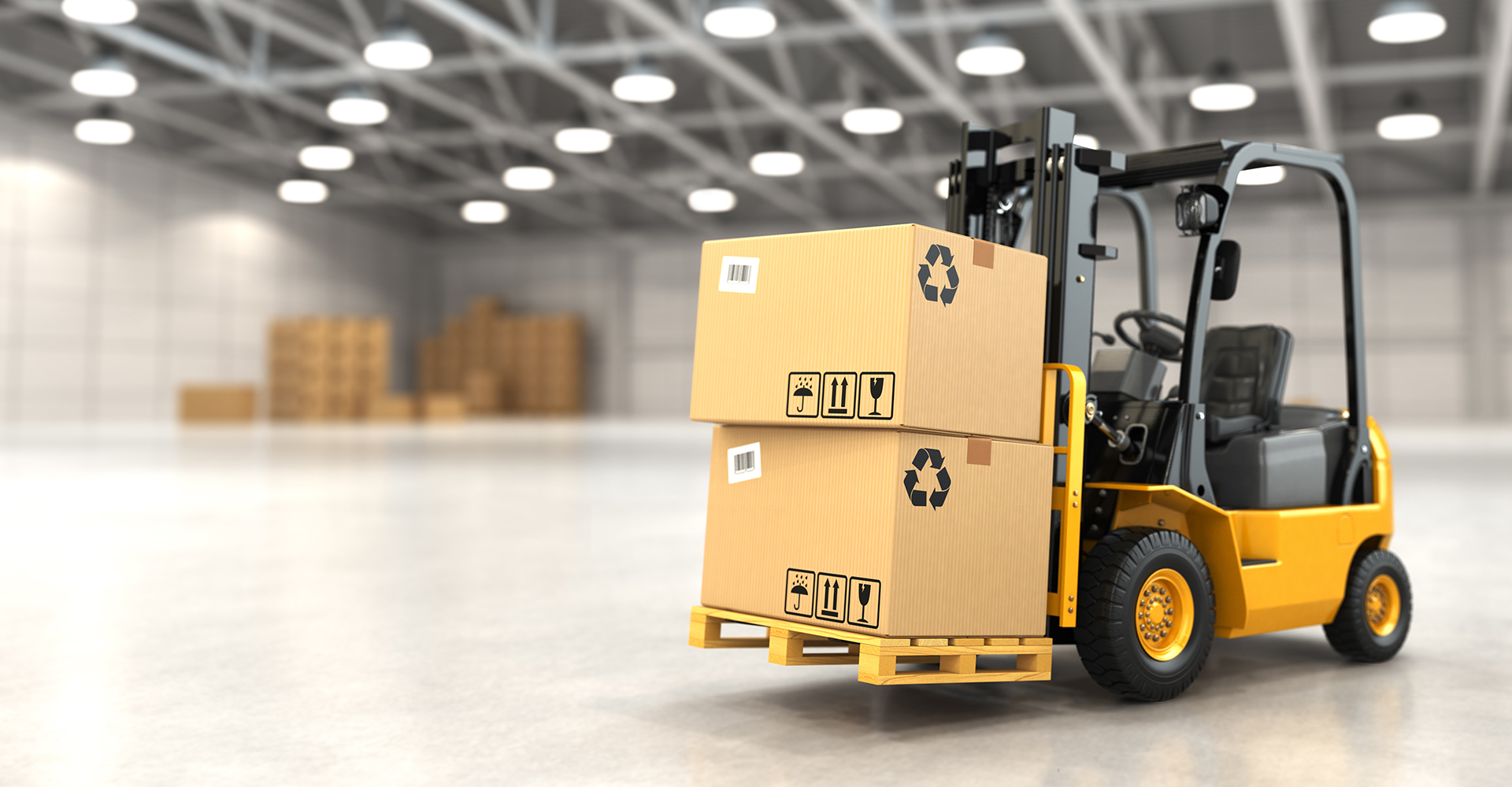
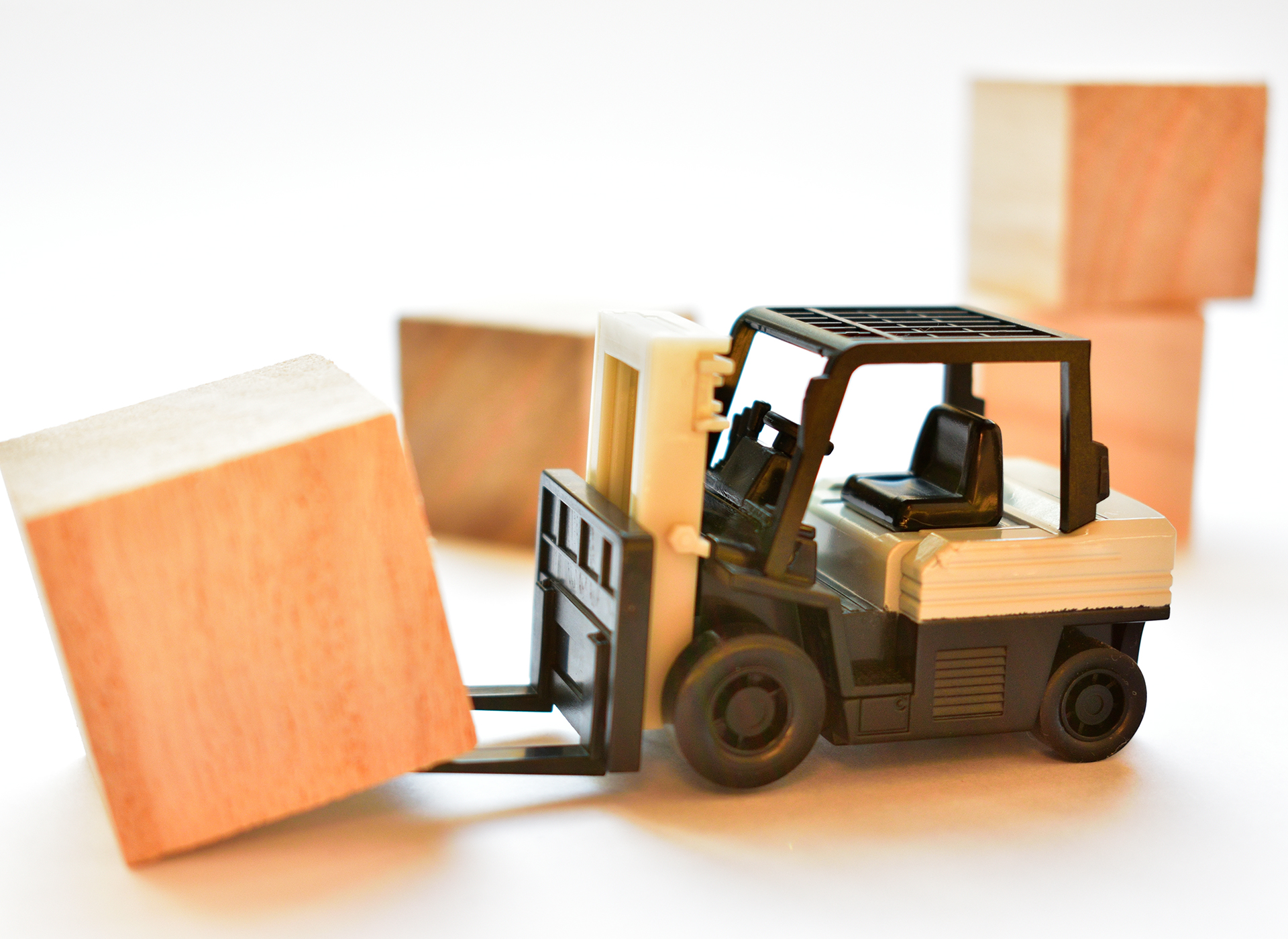
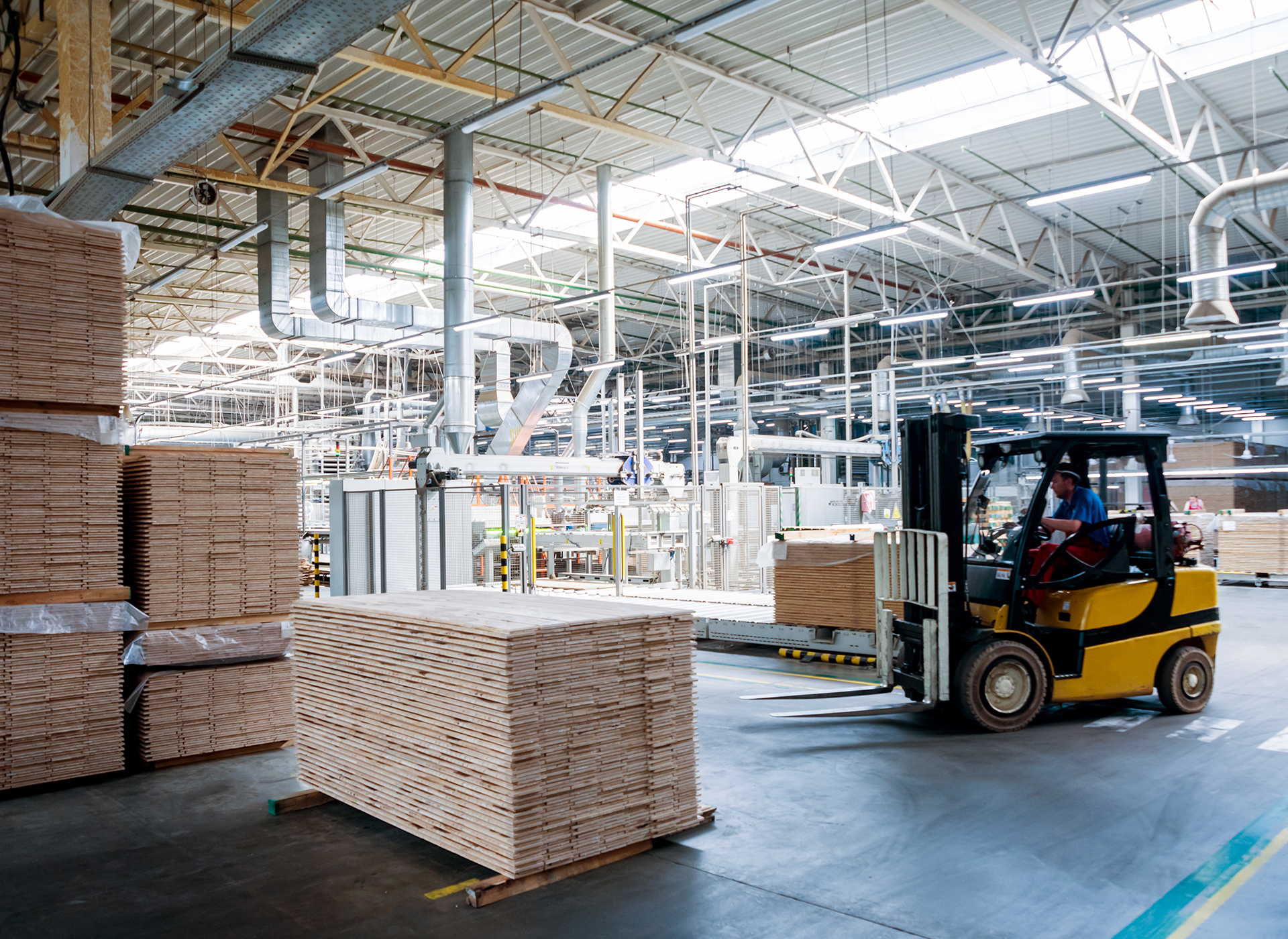
フォークリフトを使うときに必要になるのがパレットです。荷物の塊をパレットの上に積み、下の部分にフォークを入れて移動させます。フォークリフトを操作するに当たってはパレットをいかに効率よく扱えるかが大…
必要になるスキルはフォークリフト操作に関する物だけではない?あったら良いスキルとは
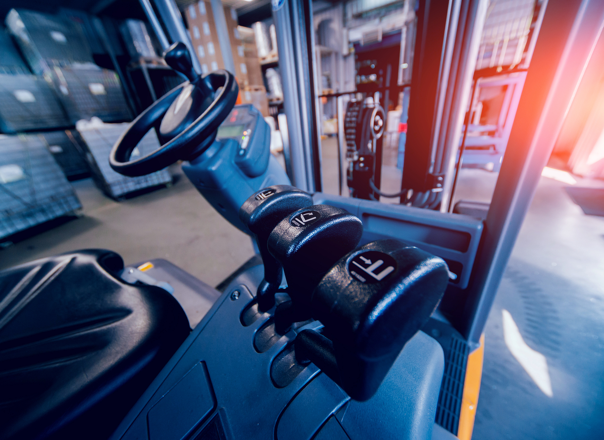
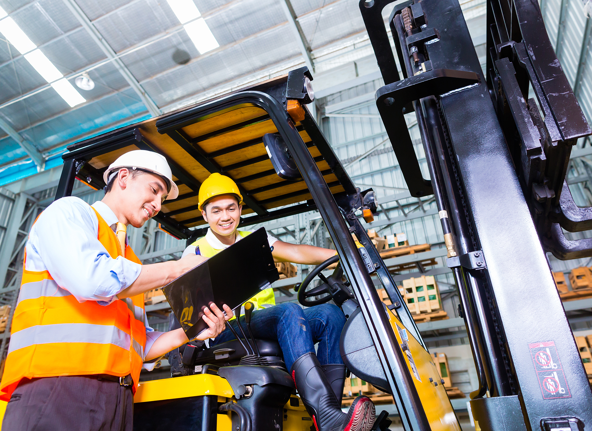
フォークリフトは、対応している免許があれば操縦そのものは誰でも可能です。ただその操縦を仕事として行うのであれば、まず安全面にしっかり気を配れるスキルが必要です。働く場所によっては狭かったり障害物の…
将来を見据えて他業種にうつる事を考えているなら?目を向けると良いスキルについて
フォークリフトの求人に応募するには資格が必要です。一般的な職場は工場内や倉庫内での作業になるので、フォークリフトに関する資格としては免許ではなく講習の修了証があれば済みます。フォークリフトのメーカ…
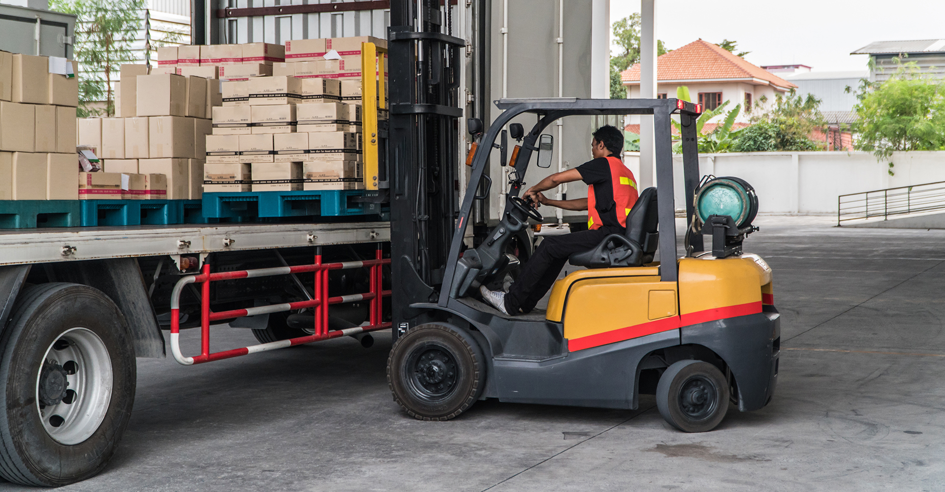
乗りっぱなしフォークリフトの求人
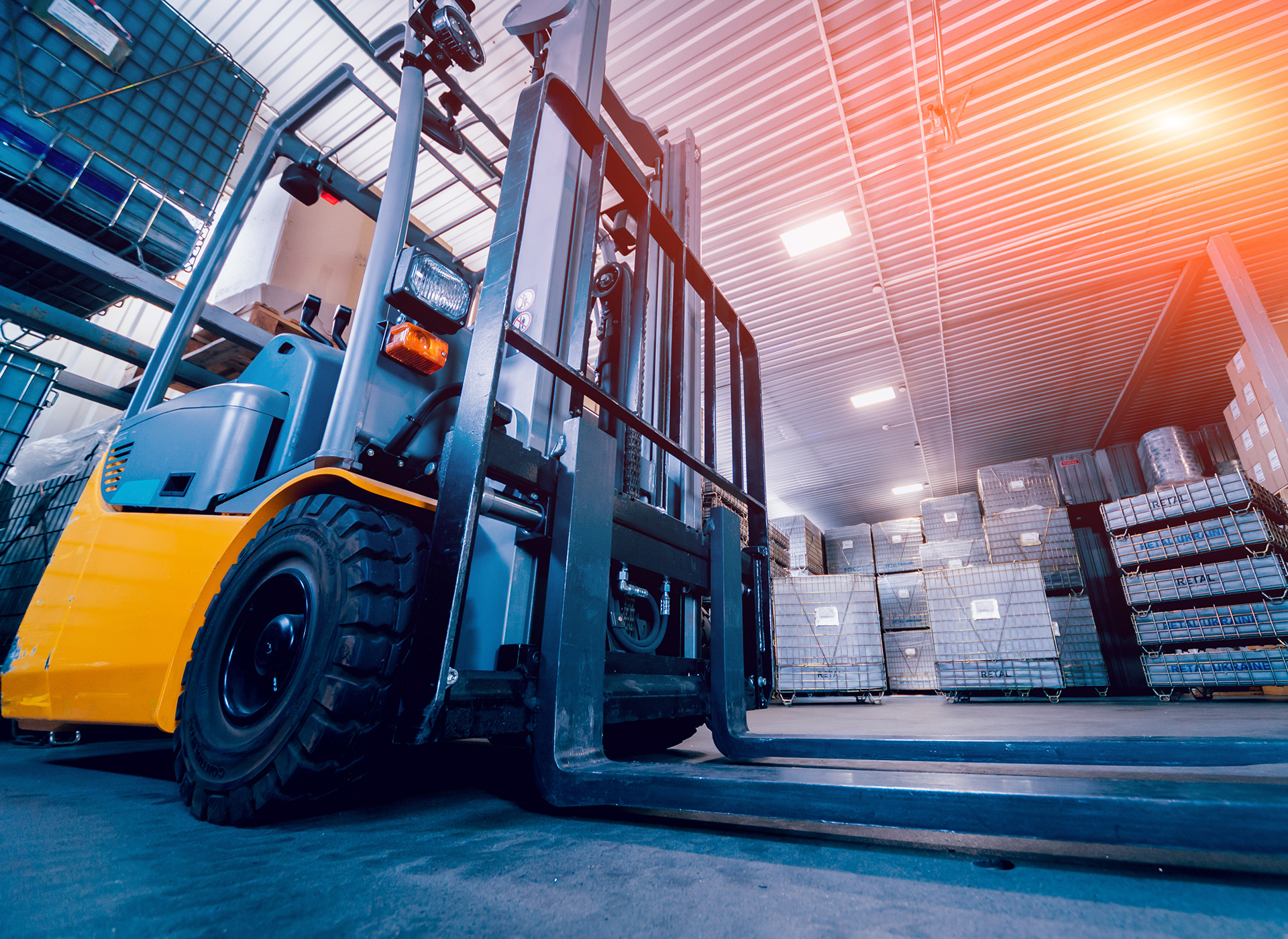
フォークリフト技能講習終了証を持っている方必見!物流センター内で入出荷作業がメインのフォークリフトの求人です。勤務中はほぼ乗りっぱなしです!
フォークリフト求人を受ける時の注意点
フォークリフト求人は、フォークリフトを操作して物を運ぶだけというイメージがあります。しかし、どんな仕事でも気をつけなければならない事が存在しています。これから、求人を探す際に知っておいた方が良い注意点について詳しく伝えていきます。
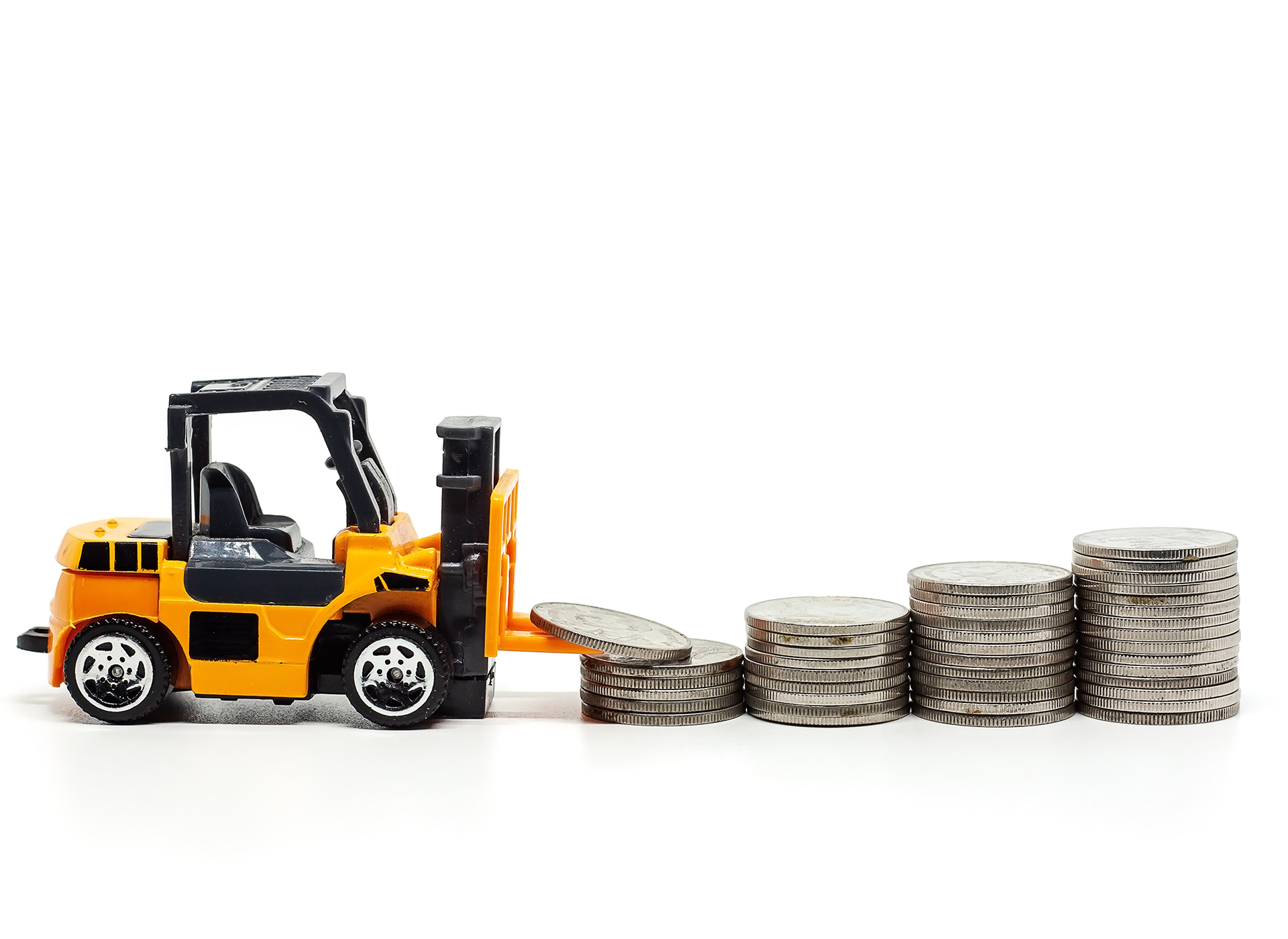
様々な求人を吟味する時に注意!給料を比べる時目を向けたいポイントとは?
求人を探す際、必ず給与はチェックすると思います。フォークリフトに関しては、運転するのに資格が必要であること、そして専門技術を要する仕事であることから、平均の求人賃金が高めに設定される傾向があります…
体を壊さないために注目したいポイント!ちょうど良い労働時間とは?
工場には機械を一切止めずに操業しているところもあれば毎日一定時間操業のところもあります。フォークリフト求人に応募するときにはその職場がどんな労働時間で働く仕組みなっているかを知っておく必要があるで…
未経験者歓迎・経験者歓迎と書かれた求人票をチェックする時の注意点
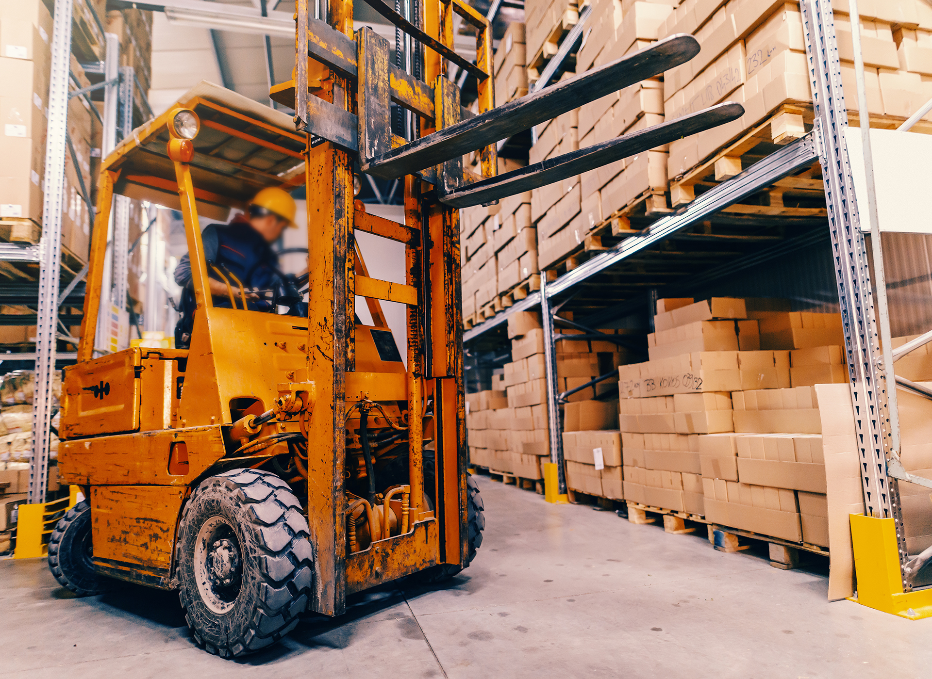
工場や倉庫などでよく走っている車両としてフォークリフトがあります。大きくて重い荷物を水平や垂直に移動できる優れものと言えるでしょう。基本的には運転技術や免許が必要ですが、求人票の中には未経験者歓迎…